


The Making of a Plastic Surgeon- Two Years in the Crucible Learning the Art and Science
Chapter 4- Jackson

Chapter 4- Jackson
“There may be people who have more talent than you do, but there’s no excuse for anyone to work harder than you do.”
Derek Jeter
For the residents assigned to the Veteran’s Administration (VA) Hospital, Victoria Hospital, and Jackson most weeks started out the same. We would meet with Dr. Millard in the Jackson cafeteria for an hour to eat breakfast and discuss the cases scheduled for that week. This was the same meeting where I had my disastrous interview months earlier. The resident working directly with Dr. Millard was at that breakfast as well, but he was usually spared the boss’ attention and could relax a little, as could the Victoria resident, while the others were in the hot seat, especially the chief resident at Jackson. He was expected to present his surgical cases for the week and be prepared to discuss and, if necessary, defend his operative plans. When there was time, the resident at the VA Hospital would do the same.
If the chief resident was lucky, the breakfast conversation might turn to one of Dr. Millard’s favorite topics- sports. Dr. Millard followed the University of Miami Hurricane football and professional boxing. A resourceful chief resident could initiate a sports-related discussion with a well-timed question about a weekend game or bout and spare himself some of the scrutiny and grilling he might otherwise have endured, but it was a ploy that had to be used sparingly. After breakfast Dr. Millard would go to the operating room with his resident. The other residents would disperse to their various rotations.
Depending on the day of the week, Raiffe and I would start the day in one of several ways. If we had surgery scheduled, we would go upstairs to our operating room to get started. Other days we would go see consults in the hospital or head over to the resident’s clinic in the outpatient clinic building. This was where service patients for nearly all the various specialties were seen. Over the first few weeks, it dawned on me that the plastic surgery residents were the de facto plastic surgery consultants for Jackson Memorial Hospital. Although Leo saw consultations, these were largely for private patients, who were a minority of the patients at Jackson. In one of the largest medical centers in the Southeast U.S.A, if you requested a plastic surgical consultation for reconstruction, you got us. Even the attending physicians on other services deferred to us on matters of reconstruction. It was a sobering realization. The consults reflected the broad scope of the specialty. We worked, literally, from the top of the head to the bottom of the feet.
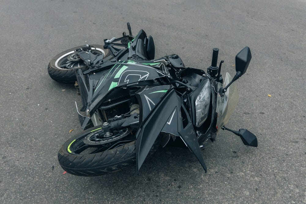
With patients like Dave on one end of the body, we had others, like Eric, on the other end. Eric was young man in his twenties who laid his Kawasaki Ninja motorcycle down on a slick Miami road one night at high speed. The orthopedic surgery service had managed a severe fracture of his lower leg between the knee and ankle. This was a high impact injury and the two bones, the tibia and fibula, were shattered into so many pieces that plates and screws could not be used. The orthopedic surgery residents had stabilized the fracture site using a device called an external fixator. They drilled large metal pins through the bones above and below the fractures then attached an external metal scaffold to them. This had articulating arms that could adjusted via universal joints, then locked in place to hold the bones in proper position for healing. The violent nature of the injury had destroyed some of the skin, fat, and muscle around the injury site leaving the fractured bones partially exposed. Without good, stable soft tissue cover over the bones, such fractures will not heal and are almost certain to eventually become infected. In the past, such injuries often required amputation. That is where we came in. Our role was to cover the fractures with healthy tissue.
The muscular bulge of the calf consists of two muscles, the soleus and gastrocnemius. The larger gastrocnemius is anatomically comprised of an inner and outer half, each with its own separate blood supply. This allows one half of the muscle to be sacrificed without compromising leg function to any significant degree. We took the inner half of Eric’s gastrocnemius, detaching it from the Achilles tendon near the ankle but left it attached to the bone at the knee, taking care to preserve its blood supply via blood vessels behind the knee joint. This allowed us to rotate the muscle over the fracture site and cover the fractured bone with good, healthy muscle. The living muscle served to bring healing factors to the fracture site to encourage bone healing, as well as antibiotics to deal with any bacterial contamination. To cover the raw muscle, we used a skin graft taken from his thigh. The fracture went on to heal, the leg was salvaged and, Eric would eventually be able to walk, even run, on it normally. To us, his horrifically scarred, but otherwise functional, leg was a thing of beauty. Eric agreed.
One final patient from those first few weeks was of interest in that his reconstruction called for techniques that dated back to the early days of the specialty when Sir Harold was reconstructing war casualties from the First World War. Dr. Millard took great interest in these cases and followed our progress closely. Paul came to us from the urology service. He was an obese young man in his thirties with diabetes who developed Fournier’s gangrene. Fournier’s is a fulminating, life-threatening infection localized to the area of the genitalia, anus, groin, and lower abdomen. It can begin with a tiny cut or scrape, or simply a pimple from an infected hair follicle. The infection is caused by several bacteria working in concert which makes each one more virulent that they would be on their own. Such infections can progress with terrifying speed; patients can go from being healthy to near death in as little as twelve hours. Patients with Fournier’s require aggressive treatment with antibiotics and surgical debridement of all infected skin and soft tissue. The latter often leaves huge open wounds that can be a challenge to close or cover.
In Eric’s case, the surgeon’s efforts saved his life but left him with no skin on the shaft of his penis or on his scrotum. His testicles lay exposed and vulnerable, looking like a pair of hideous bell clappers. Once the infection was controlled, the plastic surgery service was consulted to reconstruct his scrotum and penis. The residents who preceded us had buried the testicles under the skin of each thigh, to preserve and protect them. The raw area on the shaft of his penis they covered with skin grafts taken from his legs. At this point, his open wounds were covered and he was, strictly speaking, healed. His testicles could not, however, remain buried. Testicles must be cooler than body temperature since sperm cells cannot survive at body temperature. In addition, testicles that remain for long periods at body temperature are at higher risk of cancer years down the road. Last, but by no means least, he looked grossly disfigured. Our job was going to be to get his testicles back out where they belonged, in something resembling a scrotal sac.
We performed the reconstruction in two stages. The first involved drawing out two paddle-shaped flaps on the thigh skin, one over each buried testicle. We then cut the skin along the outline of the flaps except along the base of the flaps. The cuts went through the skin and into the fat, but no further, yet. We then stitched them up. These superficial incisions were intended to stimulate a redirection of blood flow in and out of the skin flaps, so that more blood was coming in from the base of the flaps, which would later serve as their sole point of attachment when the flaps were fully mobilized. This process of re-routing the blood flow is called delaying the flaps. After a couple of weeks, we could then fully mobilize the flaps, leaving them attached only at the narrow base, with greater assurance that they would have sufficient blood flow to survive. At the second operation we swung them, along with the testicles, under the penis and sewed them together, each flap formed half of the new scrotal pouch. In Eric’s case, there was enough loose skin on his inner thighs that we were able to simply close the donor sites with sutures. The reconstruction process incorporated several of Dr. Millard’s principles. We made a plan and a pattern for the surgery. We applied the Robin Hood principle, taking from the thigh, which had skin to spare, and transferring the skin to where it was needed. The principle of perfecting one’s craftsmanship came into play as well, to obtain a good result. Everything went smoothly. Paul made a full recovery, his testicles safely housed in their new scrotal pouch.
And so it went. Those first two months were a blur of activity with busy days seeing patients in our clinics, consultations in the hospital and from the emergency room, performing surgery, and, poring over my books in the evenings to try to assimilate as much as possible. It was stressful but at the same time exhilarating. I was reviewing anatomy I had not used since cadaver dissections in medical school. The ingenuity behind some procedures was truly amazing, especially when one considered that what we were doing had to be done by someone for the first time, with far less knowledge and fewer resources than we had.
One of the not-so-pleasant revelations of those first weeks was that the plastic surgery service shared in the management of jaw fractures. At the Naval hospital in Oakland, all jaw fractures went directly to the oral surgeons and the general surgery residents did not rotate on that service. At Jackson, responsibility for emergency coverage of facial trauma was shared by three specialties- plastic surgery, otolaryngology or ENT (ears, nose, and throat), and oral/maxillofacial surgery. Oral/maxillofacial residents were dentists who were taking additional training to learn to perform surgery of the mouth and jaws. The junior resident in plastic surgery at Jackson was therefore on call for such emergencies every third night. That was me.
I had never worked in the mouth and the surgical anatomy and techniques for treating jaw fractures were totally unfamiliar. I had to learn about normal and abnormal dental occlusion- the way the upper and lower teeth fit together. I had to learn the various types of jaw fractures and their management. Jaw fractures were a depressingly common injury in the Jackson emergency room and we could count on one most nights we were on call. The circumstances surrounding most jaw fractures were overwhelmingly limited to a few, all too typical, scenarios that we grew to accept with resignation. Nearly all involved alcohol. There was the drunk driver without a seatbelt who slammed into the steering wheel with his jaw (jaw fracture patients were nearly always men), the bar brawler who was punched in the jaw, and the drunk who stumbled and fell, leading with his chin. At Jackson, truly innocent victims with jaw fractures were the exception. So was good dental hygiene.
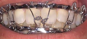
Most jaw fractures could be stabilized in the emergency room, the patients discharged and followed up in our clinic. Stabilizing jaw fractures involved wiring the upper and lower jaws together using metal bars, called arch bars, secured to the teeth with stainless steel wires. For some of the more stable fractures, this sufficed, and the jaws could be left wired together from about six weeks to allow the fracture to heal. We admitted those with unstable fractures requiring surgery. We would operate on them after a couple of days, when the inevitable swelling had subsided, making surgery easier.
The process of wiring arch bars produced dozens of sharp wire ends in the course of twisting and clipping the wires. It was common to be pricked, not once, but several times through our gloves, sometimes drawing blood. The wire tips were so sharp that even double-gloving (wearing one glove over another) could not always prevent these punctures. More than anything else in the residency, management of jaw fractures brought up the specter of Acquired Immunodeficiency Syndrome (AIDS).
AIDS arrived on the scene in early 1980s, when I was still a general surgery resident. At the time, the cause was unknown. We were often asked to place intravenous lines in young, gay men who looked terribly gaunt and were desperately sick. The diagnosis in the medical record was usually listed simply as Gay Related Immune Deficiency Syndrome or GRIDS. By 1987, we knew the cause was a virus but there was still much we did not know. Reputable authorities were still concerned that AIDS could be spread by casual contact and possibly even by such environmental exposures as mosquito bites, a big concern in a mosquito-ridden state like Florida.
One big worry for residents at Jackson was the prevalence of HIV/AIDS in the patient population seen in the emergency room. Estimates ran as high as ten percent. Obtaining a blood test for HIV/AIDS required that the patient sign an informed consent on par with that required for major surgery. The patient’s privacy trumped the fact that a physician or other health care worker had been stuck with a contaminated needle. Many patients refused testing because of the stigma attached to having AIDS at that time. Testing then was somewhat academic in any event since there was no effective treatment for HIV/AIDS. Drug therapy was still in its infancy. Acquiring a chronic illness, such as hepatitis, from a patient had always been a risk of practicing medicine, but AIDs now presented us with the possibility of acquiring an incurable disease that was also usually fatal.
This was dramatically impressed on me just after returning to Miami. While looking for a place to rent, we visited a townhouse that was being vacated by a resident who had just completed his training at Jackson. Weeks earlier he had been accidently stuck with a needle contaminated with blood from an AIDS patient and developed an acute case of HIV infection. As we spoke to his wife, his two young children were playing in another room, and he was lying on the sofa, too sick to get up. The impact this very common hospital occurrence would have on this young family was incalculable. It certainly made an impression on me. I thought about that resident many times as I placed arch bars in jaw fractures in the emergency room. Jaw fractures were an area of plastic surgery that I never warmed to, and it was with relief and no regrets that I gave up treating these altogether years later, when I was in practice for myself.
As a plastic surgery resident, I saw a different side of burn care. I had spent time on a burn unit in San Francisco, treating acute burns. The priorities were to save life, preserve function, and restore normal appearance, in that order. Raiffe and I did a weekly walk through of the Jackson burn unit with the doctors and nurses there to see which patients might be ready for reconstruction.
Burn patients are among the most tragic and difficult to care for in any area of medicine and acute burn care is one of the most demanding areas of medical/surgical practice. Severely burned patients are desperately sick and just getting them to where reconstruction comes into play can be a daunting challenge. Burns are incredibly destructive of soft tissues and burn scars are particularly prone to complications. Burn scars often become thickened even to the point of producing painful, disfiguring masses of scar tissue called keloid scars. Burn scars over across joints can contract, bending the joint so it cannot straighten and nothing short of an operation will correct this. On the face, contracted scars can hideously distort normal features. Maria was one such patient.
Maria was a beautiful 3-year-old Hispanic girl. Her cherubic face had huge, dark eyes, framed by thick ringlets of black hair. Her features were perfect. At least, they had been before she was burned. Maria and her 5-year-old brother were playing in a closet in their home. He had some matches. The closet caught fire. Her brother escaped with minor burns, but Maria did not, her face severely burned. By the time we saw her, her burns were essentially healed, but looked hideous. There was a hole where her nose should have been. Her eyelids were pulled apart so she could not close her eyes, which had to be lubricated continuously to avoid ulcers and blindness. Her lips were pulled into a snarl-like rictus. The heartbreak of her situation was impressed upon us every time we saw her because of a photograph above her bed, showing her before the fire.
Raiffe and I began her reconstruction by releasing the contracted scars on her eyelids and placing full thickness skin grafts taken from her genital area, where the thin skin there best matches the thin skin of the eyelids. This was Millard’s principle of replacing missing tissue with tissue as much like it as possible. In contrast to the thin, partial thickness skin grafts used in early burn care, full thickness grafts would not contract and this would allow her to open and close her eyes normally. We also began the process of moving unburned skin from here shoulders up to her face to replace the burn scars around her mouth. For that, we turned to tissue expansion.
Maria was my introduction to tissue expansion. If stretched slowly, skin has an almost unlimited ability to expand, as evidenced by people whose weight increases to astronomical heights of 500 pounds and beyond. If you stress skin gradually, you stimulate the skin to grow producing potentially unlimited amounts of skin that can be used for reconstruction. The first case of tissue expansion was presented at a plastic surgery meeting in Miami Beach in 1956 by Dr. Charles Neumann, a plastic surgeon from New York. He placed a balloon under the skin adjacent to the ear of a 45-year-old man who had lost the upper half of his ear to trauma. He gradually inflated the balloon over a period of two months by means of a tube that he tunneled down to the neck, producing enough extra skin to successfully reconstruct the ear. Surprisingly, Neumann’s presentation and subsequent paper were received with a collective yawn by the plastic surgery community, and it was not until 1975 that tissue expansion was re-discovered by Dr. Chedomir Radovan, a plastic surgeon at Georgetown University. Radovan developed the modern tissue expander that came to bear his name and it was through his efforts to promote the process that tissue expansion was finally accepted by plastic surgeons and became an integral part of the specialty. Radovan died tragically at the young age of 52 when he drowned while swimming in the ocean off Miami Beach.
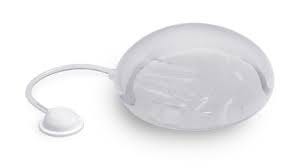
For Maria, in addition to the full thickness skin grafts on her eyelids, we also placed a pair of tissue expanders under the unburned skin of her shoulders. These could be accessed via injection ports, connected to the expanders by a tube and tunneled under the skin a distance away. With early expanders, the port was left outside the skin for easy access to it, but these had a higher rate of infection, so the ports were eventually buried under the skin as well. Current expanders have the injection port built into the expander itself, eliminating the tube and separate port. Once our expanders were in place, we could inflate them by injecting sterile saline through the port on a weekly basis.
As the expander grew in size, the constant tension created under the skin stimulated skin growth. Beyond the needle prick, the expansion process was essentially painless. Most patients undergoing tissue expansion can go about their daily affairs with no real restrictions.
With Maria, we knew we would not be finishing this in time to perform her next surgery before we moved on. It would be up to later residents to use this expanded skin to replace some of the burn scars on her lower face and around her mouth. Maria was going to be an ongoing project for several future generations of residents.
Of course, as this was a plastic surgery residency, we also performed cosmetic surgery. The juxtaposition of patients such as Juan, Eric, Paul, and Maria with our cosmetic patients made for a truly discordant mix. At Jackson, nothing was done in moderation, and this carried over to the cosmetic surgery aspect of our training.

If you want to read ahead rather than wait for additional chapters to be posted, you can order the book on Amazon as an eBook or paperback.